Biological/Environmental Indicators and Responses (BEIR)

Biological/Environmental Indicators and Responses (BEIR)

The Biological/Environmental Indicators and Responses (BEIR) Facility Core provides CEED investigators and trainees with ready access to cutting-edge technology for analysis of indicators of exposure and biological responses to established and emerging environmental toxicants and particles in humans, animals, cells, and tissue models. Biological indicators and responses include changes in cell phenotype, function, protein expression, metabolism, oxidative stress, and structure.

The Core works closely with the Translational Research Support Core (TRSC), the Human and Animal Exposure (HUMANE) Facility Core, and the other CEED Research Cores on optimization of experimental design for collection of samples from animals and human participants, scientific rigor, and appropriate technology/instrumentation, to  ensure the highest quality data.

Core Director

Photo of Debra Laskin PhD
Debra Laskin, PhD
CEED Deputy Director Rutgers-EMSOPPharmacology and Toxicology

BEIR Core Advisory Group

Photo of Lauren Aleksunes PharmD, PhD
Lauren Aleksunes, PharmD, PhD
Assistant Professor Rutgers-EMSOPPharmacology and Toxicology
Photo of Alison Bernstein PhD
Alison Bernstein, PhD
Associate Director
Photo of Andrew Gow PhD
Andrew Gow, PhD
Rutgers-EMSOPPharmacology and Toxicology
Photo of Jeffrey Laskin PhD
Jeffrey Laskin, PhD
Rutgers-SPHEnvironmental and Occupational Health

Core Mission

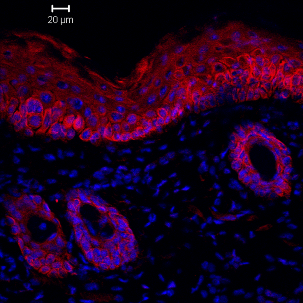
The major goal of the BEIR Facility Core is to assist CEED investigators in identifying biological indicators and responses to environmental/occupational exposures that are linked to adverse health outcomes. Results from these investigations will aid in identification of targets for the development of approaches to mitigating or limiting toxicity and disease pathogenesis.

Technology Services

This technology enables the characterization and separation of cells or particles suspended in fluid using size (forward scatter), complexity (side scatter), viability, and fluorescence.

Using lasers, this microscopy approach allows high optical resolution by acquiring fluorescent images through a spatial pinhole. Multiple images can be captured in two dimensions and then reconstructed to provide three-dimensional structures of a tissue section or cells.

Time-lapse microscopy of live cells provides investigators with an opportunity to evaluate biological responses (e.g., migration, generation of reactive oxygen species, apoptosis, ) in a spatiotemporal manner.

Multiple options are available for CEED investigators to capture spatial distribution of cells or molecular markers within tissue sections following staining with traditional  histology  stains  (i.e.,  H&E  sections)  or fluorescent markers (i.e., targeted antibodies). Available software is then used for quantification.

Metabolic activity and ATP production have emerged as central not only to phenotypic and functional activation of immune cells responding to injury and infection, but also to epithelial cells and cancer cells.

Cellular responses to a toxicant are rarely homogenous. Traditional protein-based quantification methods fail to capture this heterogeneity within a population.

Recent Core Publications

Gowdy KM, Laskin DL. Resolution of inflammation in xenobiotic-induced mucosal injury and chronic disease. Toxicol Appl Pharmacol. 2023 May 1;466:116455. doi: 10.1016/j.taap.2023.116455. Epub 2023 Mar 11. PMID: 36907382.

Pappas G, Wilkinson ML, Gow AJ. Nitric oxide regulation of cellular metabolism: Adaptive tuning of cellular energy. Nitric Oxide. 2023 Feb 1;131:8-17. doi: 10.1016/j.niox.2022.11.006. Epub 2022 Dec 5. PMID: 36470373; PMCID: PMC9839556.

Herbert J, Kelty JS, Laskin JD, Laskin DL, Gow AJ. Menthol flavoring in e-cigarette condensate causes pulmonary dysfunction and cytotoxicity in precision cut lung slices. Am J Physiol Lung Cell Mol Physiol. 2023 Mar 1;324(3):L345-L357. doi: 10.1152/ajplung.00222.2022. Epub 2023 Jan 24. PMID: 36692165; PMCID: PMC10026991.

Contact

Administrators: Jessica Cervelli & Rita Hahn

cervelli@phatmacy.rutgers.edu

rhahn@eohsi.rutgers.edu


Copyright © 2023 – Rutgers, The State University of New Jersey